
Full-chain Independent R&D
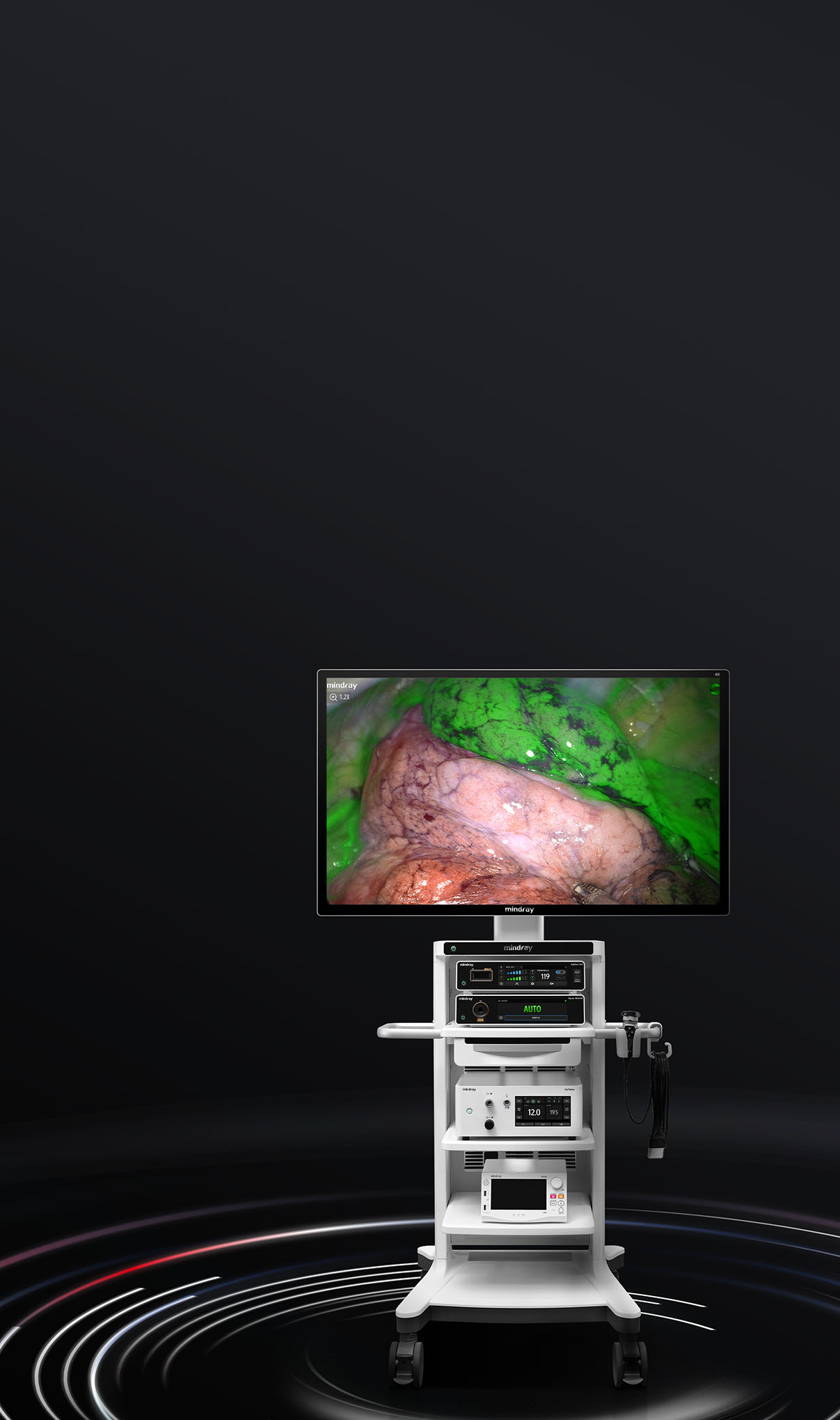
The integration of the new generation hardware platform with novel software algorithms greatly elevates image performance. AutoFocus, Automatic Scene Recognition and the Intelligent Image Algorithm work together to present high quality images. Breakthroughs in fluorescence imaging technology make hidden lesions and stained boundaries truly visible, allowing for more accurate surgical navigation.
The UX series, based on full-chain R&D, is quickly becoming a platform capable of further evolution, which holds great promise for value creation and more possibilities.

Light Source

Rigid Endoscope

Camera Head

Image Algorithm

Customized Monitor

4K Recording
Full-chain Independent R&D
The integration of the new generation hardware platform with novel software algorithms greatly elevates image performance. AutoFocus, Automatic Scene Recognition and the Intelligent Image Algorithm work together to present high quality images. Breakthroughs in fluorescence imaging technology make hidden lesions and stained boundaries truly visible, allowing for more accurate surgical navigation.
The UX series, based on full-chain R&D, is quickly becoming a platform capable of further evolution,which holds great promise for value creation and more possibilities.


Smart ViewExceptional Image Quality
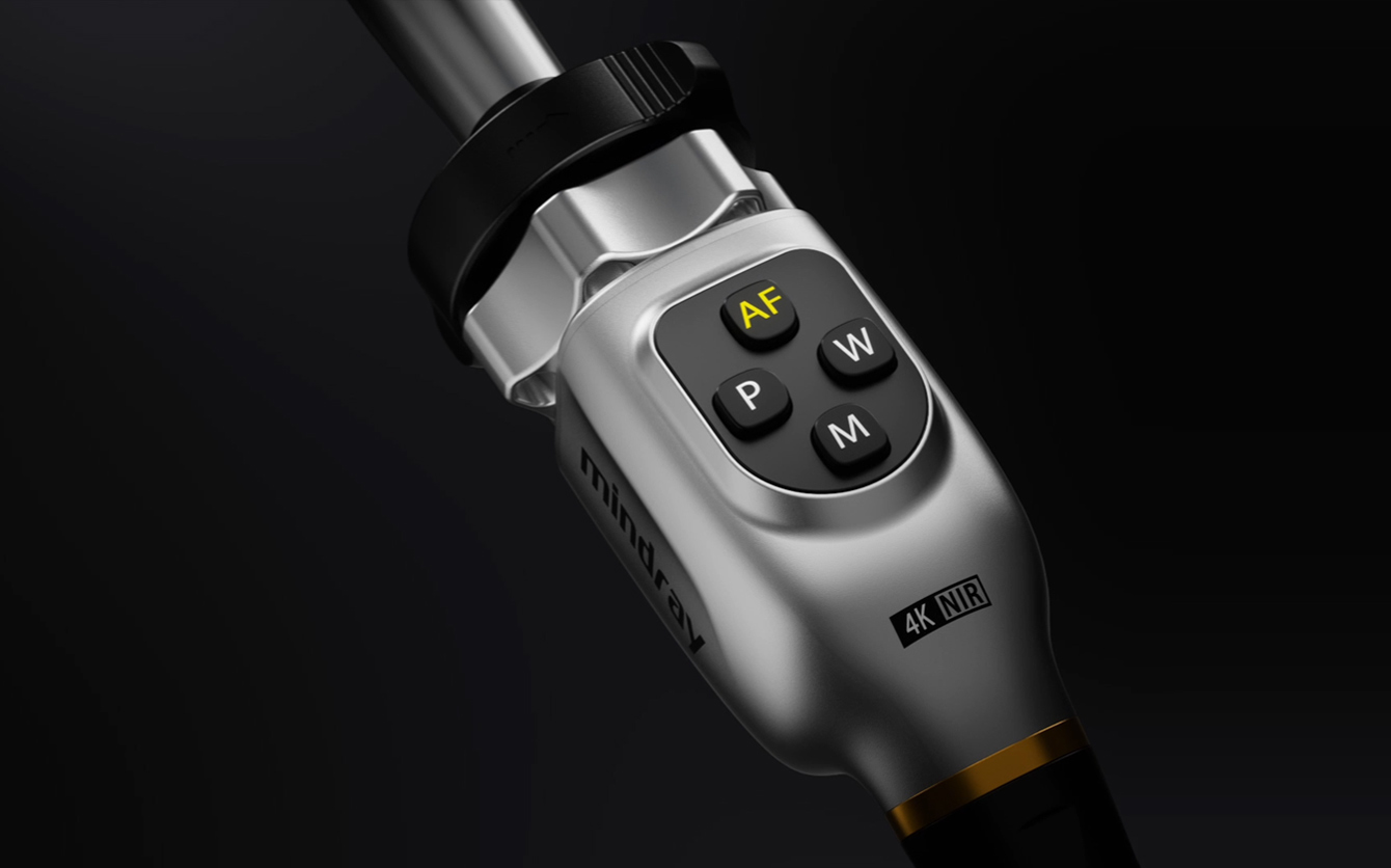
AutoFocus, and high quality image achieved with one touch
The perfect combination of mechanical structure, optical design, and the powerful computing power of the new imaging chip brings effective precision focusing and reduces manual operations, allowing the surgeon to focus on the surgery with a perfect image.
Smart View
Exceptional Image Quality
AutoFocus, and high quality image achieved with one touch
The perfect combination of mechanical structure, optical design, and the powerful computing power of the new imaging chip brings effective precision focusing and reduces manual operations, allowing the surgeon to focus on the surgery with a perfect image.
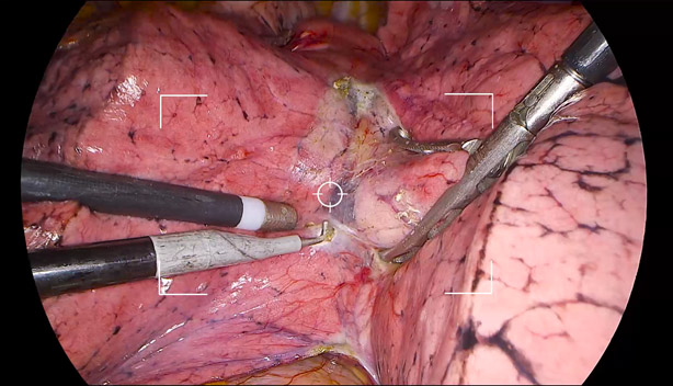
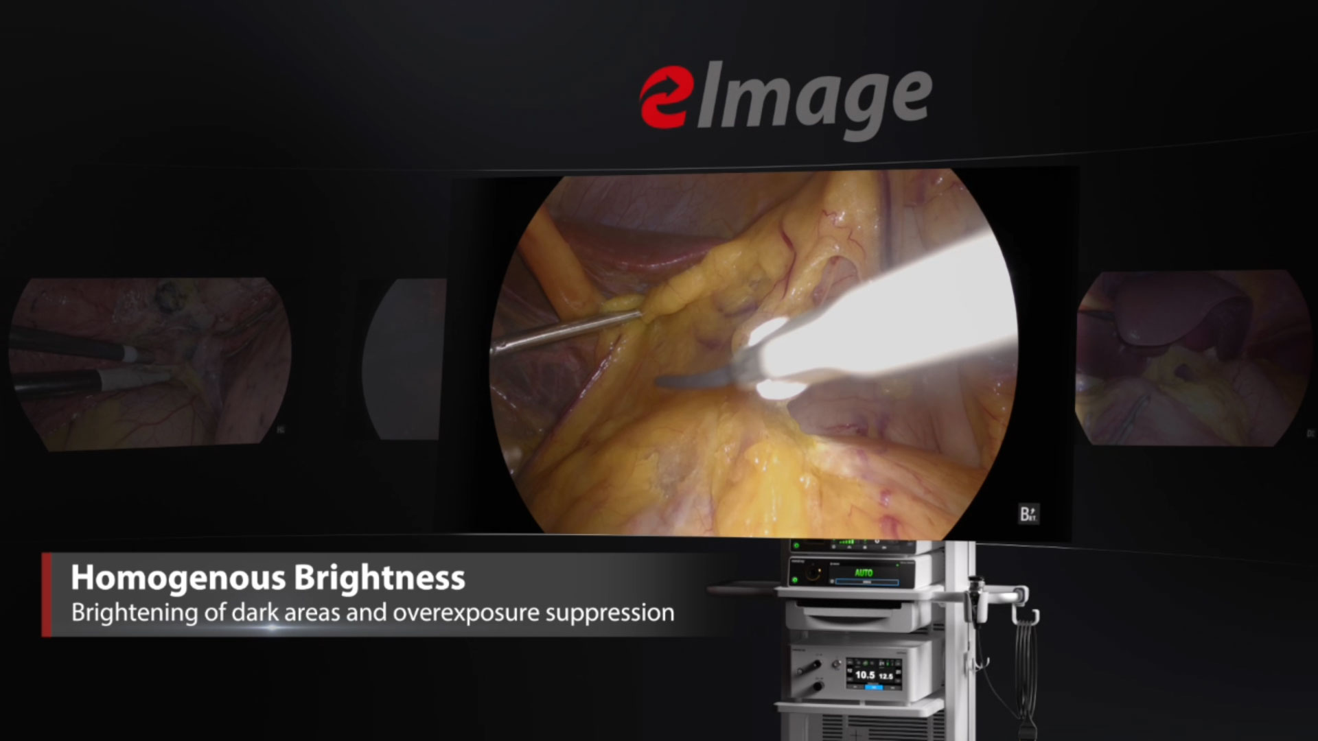
Automatic scene recognition, intelligent brightness adjustment
Smart exposure: Determine different detection areas according to different scene and accurately match the exposure parameters without the need to manually switch department modes.

Small diameter scope scene (e.g. hysteroscope)

Laparoscope scene
Automatic dimming: The camera system can automatically adjust the intensity of the light source in real time according to the exposure requirements of the current image, and ensure appropriate brightness.
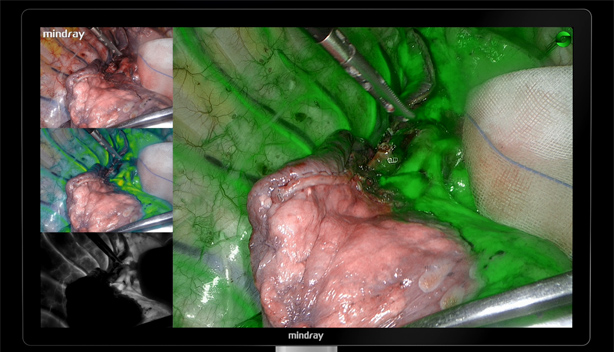
 Intelligent Image Algorithm, Even in Extreme Circumstances
Sensitive perceptionPrecise Navigation
 The breakthrough in fluorescence technology significantly boosts detection sensitivity
and fluorescence imaging stability, leading to more precise navigation.
The breakthrough in fluorescence technology significantly boosts detection sensitivity
and fluorescence imaging stability, leading to more precise navigation.
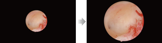
Ultra-high fluorescence sensitivity
Dual optimization of excitation and imaging brings the fluorescence signal capture sensitivity down to the nmol level, which helps the clinical detection of small metastatic lesion at low doses and delivers greater penetration capability at the same dose.
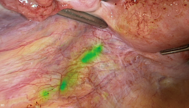
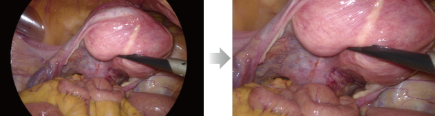
Fluorescence stabilization algorithm
Accurately displaying the ICG imaging area, effectively avoiding signal attenuation caused by distance and angle deviation, ensuring great fluorescence stability and boundary division consistency.
Fluorescence pixel-level fusion
Strict control of the assembly process helps achieve the white light and fluorescence image pixel-by-pixel alignment and fusion. The fluorescence image with white light texture details can also help with the entire fluorescence-guided surgical process.
Sensitive Perception
Precise Navigation
The breakthrough in fluorescence technology significantly boosts detection sensitivity and
fluorescence imaging stability, leading to more precise navigation.
Ultra-high fluorescence sensitivity
Dual optimization of excitation and imaging brings the fluorescence signal capture sensitivity down to the nmol level, which helps the clinical detection of small metastatic lesion at low doses and delivers greater penetration capability at the same dose.
Fluorescence stabilization algorithm
Accurately displaying the ICG imaging area, effectively avoiding signal attenuation caused by distance and angle deviation, ensuring great fluorescence stability and boundary division consistency.
Fluorescence pixel-level fusion
Strict control of the assembly process helps achieve the white light and fluorescence image pixel-by-pixel alignment and fusion. The fluorescence image with white light texture details can also help with the entire fluorescence-guided surgical process.
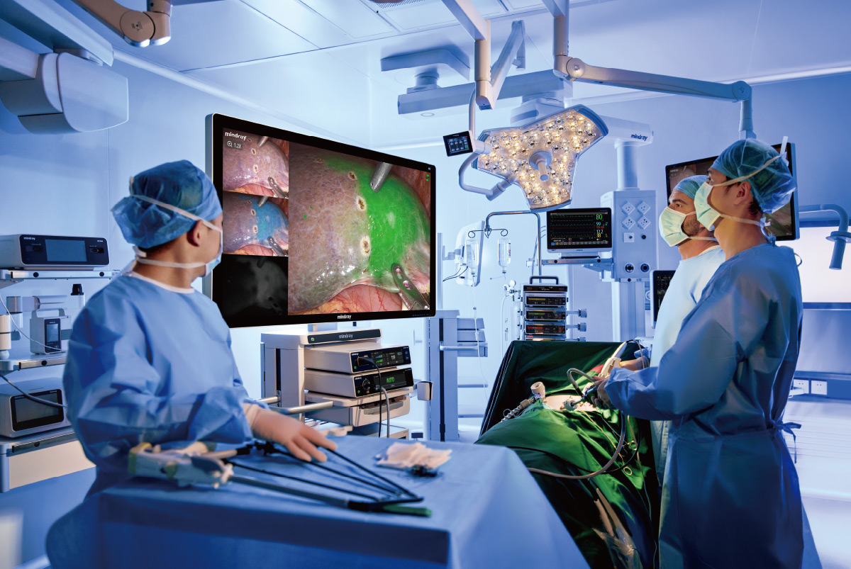
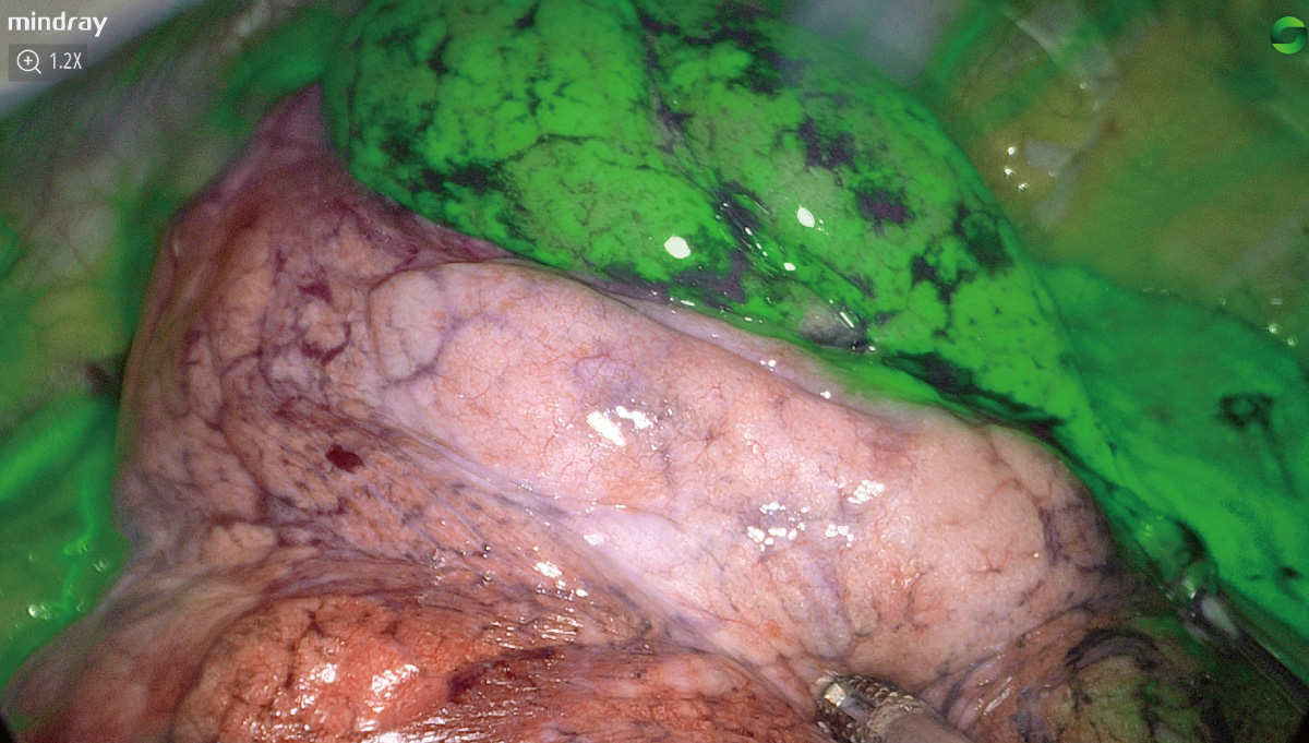
Clinical Cases
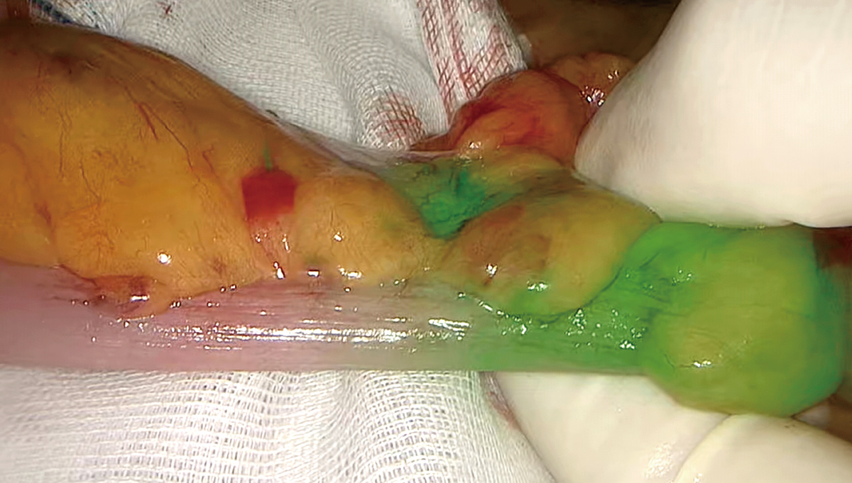
Evaluation of colorectal anastomotic blood supply

Laparoscopic liver watershed resection

SLN mapping in endometrial cancer
Anatomic subpulmonary lobe resection
Evaluation of colorectal anastomotic blood supply
Laparoscopic liver watershed resection
SLN mapping in endometrial cancer
Anatomic subpulmonary lobe resection
Intelligent ControlFlexible Mastery
Flexible Mastery
Automatic Adaptive Zoom achieved with one touch
Intelligent recognition of endoscope type and automatic adjustment with adaptive zoom reduces the need for repeated manual adjustment, and ensures ideal visibility for different surgeries.
Small diameter scope - One Touch to adaptive Zoom
Laparoscope - One Touch to Full Screen
Built-in 4K recording with Variable BitRate + H.265 encoding
4K high-quality video recording vividly reproduces the whole process of surgery, bringing quality academic sharing. Meanwhile, Variable BitRate and H.265 encoding reduce the file size of the same quality by 50%, meaning less worry about storage.

Master Control Trolley
Start all devices in the trolley with one touch

32/55 inch 4K 3D Monitor

White Light / Fluorescence Camera Head
Multiple focal lengths available
White light camera head weight: 190 g
Fluorescence camera head weight: 240 g

White Light / Fluorescence Light Source
Automatic dimming of the whole series

Medical Digital Video Recorder
Access to hospital PACS system Simultaneous recording of two signal sources available

10/5 mm Rigid Endoscope
Multi-specification fluorescence rigid endoscopes available
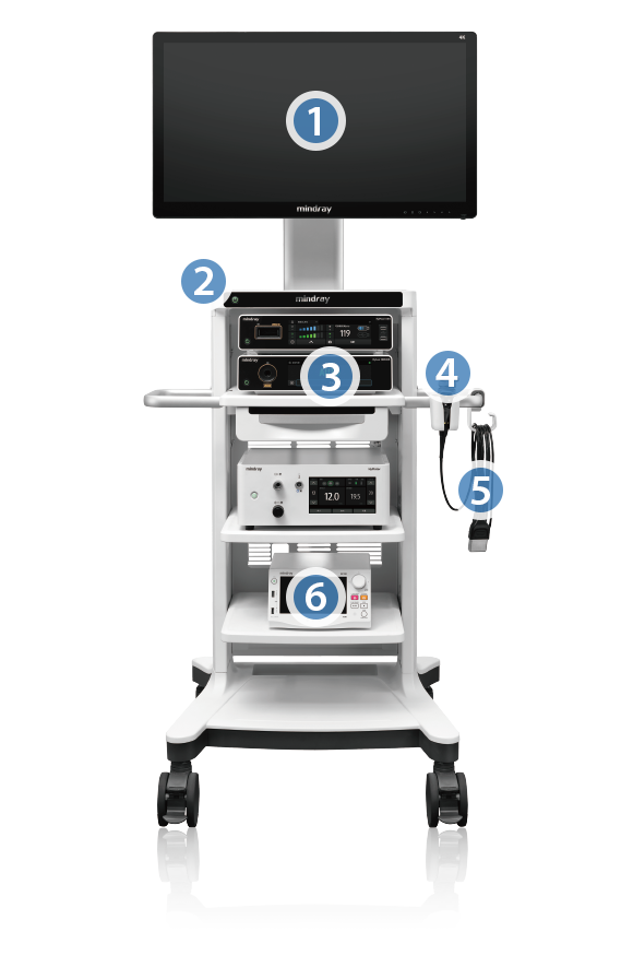
1
32/55 inch 4K 3D Monitor
2
Master Control Trolley
Start all devices in the trolley with one touch
3
White Light / Fluorescence Light Source
Automatic dimming of the whole series
4
White Light / Fluorescence Camera Head
Multiple focal lengths available
White light camera head weight: 190 g
Fluorescence camera head weight: 240 g
5
10/5 mm Rigid Endoscope
Multi-specification fluorescence rigid endoscopes available
6
Medical Digital Video Recorder
Access to hospital PACS system Simultaneous recording of two signal sources available
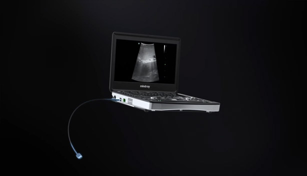
Smooth ConnectivityUnlimited Possibilities
Unlimited Possibilities
Innovative Endoscope and Ultrasound Fusion Solution for MIS
Ultrasound and Endoscopy images can be displayed on the same screen with the same brightness, and this combination of images can be expanded to multiple screens, eliminating the need for the surgeon to look back and forth for cross-referencing, helping to localize the lesion and perform accurate puncture.
Simultaneous recording on the same screen eliminates the need for surgeons to synthesize two videos by aligning timelines, making teaching and sharing more efficient.
Digital Operating Room, Upgraded Management
The UX5 Endoscope Camera System can be integrated into the Mindray digital operating room for centralized information management, enhancing the overall quality and efficiency of surgical procedures.

Digital Operating Room, Upgraded Management
The UX5 Endoscope Camera System can be integrated into the Mindray digital operating room for centralized information management, enhancing the overall quality and efficiency of surgical procedures.