Light Source

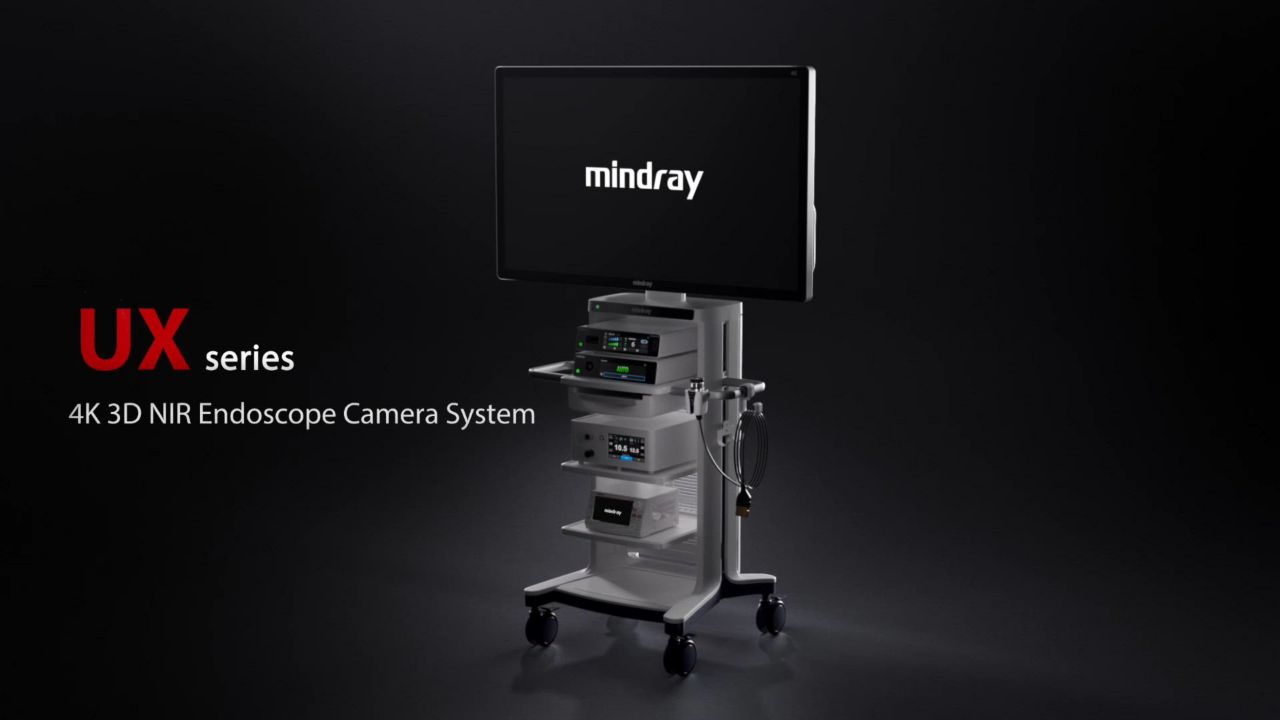
Endoscope Camera System
UX7 Series 4K&NIR&3D
Vision Beyond Imagination
Watch Product Video
Full-chain Independent R&D
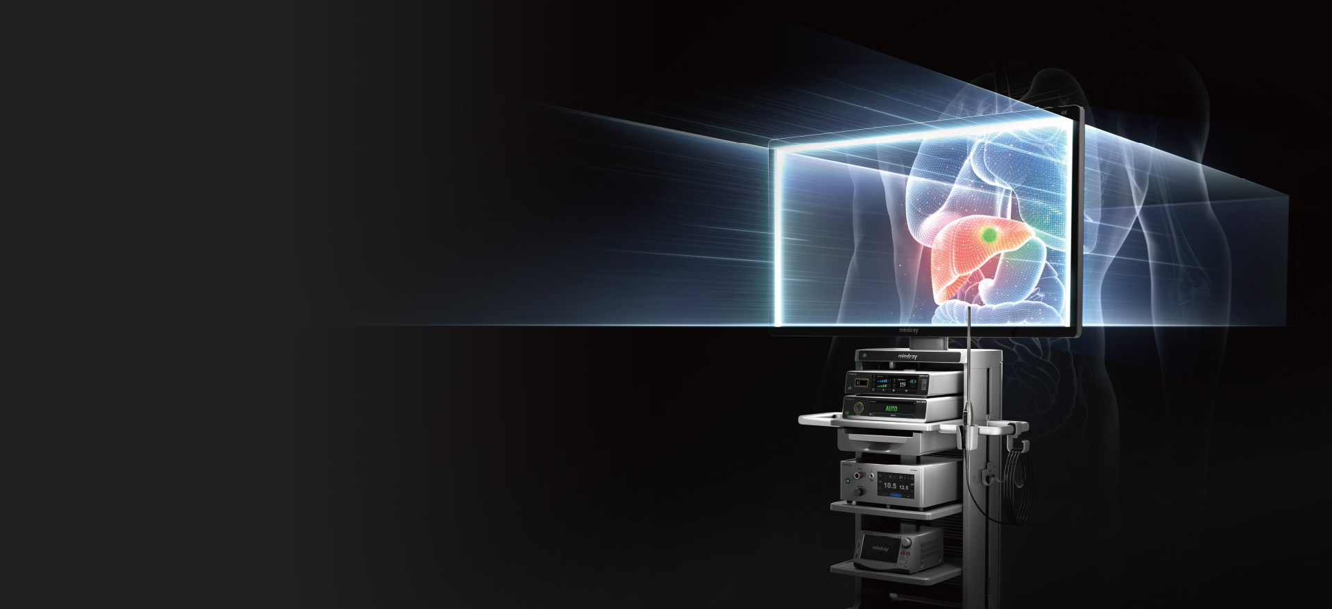
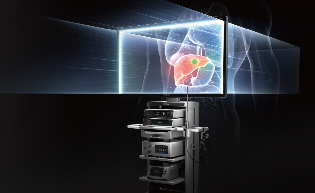
The advancement of minimally invasive technology has significantly generates the demand for precision surgeries. This has led to increased requirements for the resolution, real-time navigation, and accurate restoration of 3D structures within the cavity.
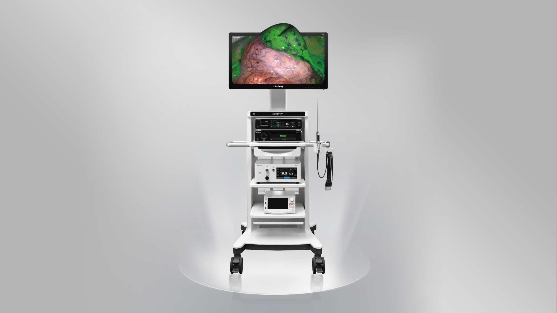
Based on full-chain independent R&D, Mindray's new generation UX Series Endoscope Camera System has been innovatively integrated with 4K fluorescence and 3D technologies. It provides a comprehensive enhancement in imaging performance and operational experience, enabling clinical breakthroughs in complex surgical procedures.
With various configuration options suitable for multiple departments, it is a truly "all in one imaging platform" for the operating room.


Rigid Endoscope

Sensor

Image Algorithm

Customized Monitor

4K Recording

{{data.title}}
{{item}}
{{data.id}}
3D Intelligent Bionics,
Natural Space Perception
Dual Chips True 4K
Dual-4K 3D imaging reproduces the structure in the cavity, making the surgery safer and more efficient.
5.6 mm Large Pupil Distance
Enhanced depth of 3D vision can discern small depth differences for more precise surgical positioning.
3D Fluorescence Imaging
Stable stereo fluorescence navigation makes surgical operations more precise.


AutoRotate Correction
Built-in high-precision attitude sensor secures real-time
perception of the endoscope movement
AutoRotate Correction of the endoscope enables all-around view
observation

Autoclave Available
A new breakthrough in sterilization processes with a high
reliability
The whole Video Endoscope supports Autoclave/Low Temperature
Plasma/EO Sterilization

Enhanced Anti-Fog
The Chip-on-Tip design achieves active heating and defogging, dramatically reducing the frequency of intraoperative lens wipes

Weighs only 420 g
Titanium alloy handle, sturdy yet lightweight, allowing for easy maneuverability

Focus-Free Design
The large depth of field eliminates the need to focus, while presenting clear image for a smoother surgical process
Smart View
Exceptional Lmage Quality
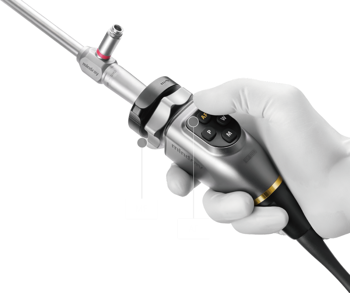
Integrated Auto And Manual Focus
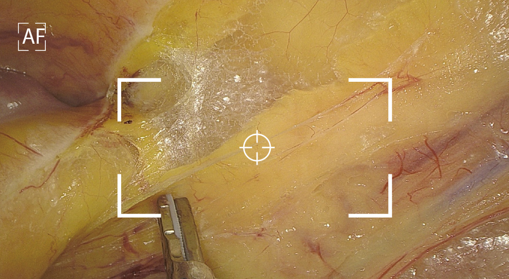
AutoFocus and high quality image achieved with one touch
Automatic Scene Recognition, Intelligent Brightness Adjustment
Smart exposure: Determine different detection areas according to different scene and accurately match the exposure parameters without the need to manually switch department modes.

Small diameter scope scene (e.g. hysteroscope)

Laparoscope scene

Intelligent Image Algorithm, Even in Extreme Circumstances
{{data.title}}
Sensitive Perception, Precise Navigation

The Fluorescence Technology Significantly Boosts Detection Sensitivity and Fluorescence Imaging Stability, Leading to More Precise Navigation
Ultra-high Fluorescence Sensitivity
Dual optimization of the excitation and imaging pathways achieves fluorescence signal capture sensitivity as low as nmol levels, which helps the clinical detection of low-dose micro-metastases and offers greater penetration capability at equivalent doses.

eFlo Stabilization Algorithm
Accurately reproduces the distribution of the contrast agent, effectively avoiding signal attenuation caused by distance and angle variations. This significantly enhances fluorescence stability, ensuring consistent boundary delineation.

Fluorescence Pixel-level Fusion
Strict control of the assembly process ensures the white light and fluorescence image pixel-by-pixel alignment and fusion. The fluorescence image with white light texture details can also help with the entire fluorescence-guided surgical process.
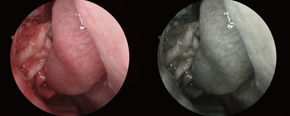
Tone Enhancement
Tone filtering is used to see through the mucosal vascular network for the differentiation of anomalous vessels to assist in clinical diagnosis.
Clinical Cases
{{data.title}}
MIS Ecosystem
Unlimited Possibilities
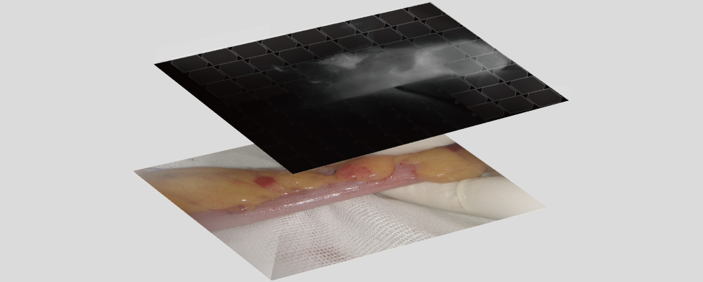
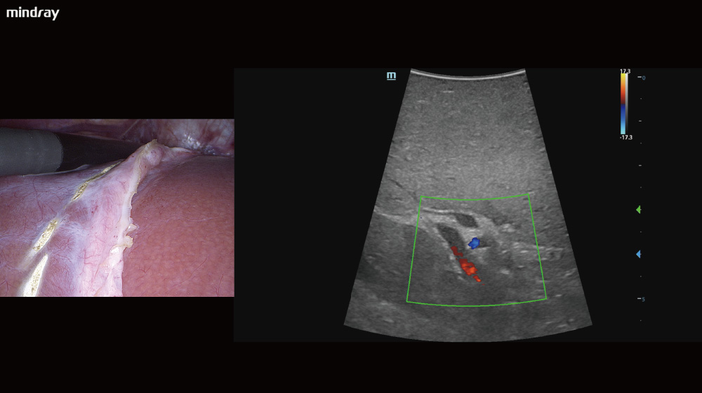
Multimodal Image Fusion
The real-time display of ultrasound images on the endoscope screen helps the surgeon localize the hidden lesion. Simultaneous recording of ultrasound and endoscope images on the same screen makes teaching and sharing more efficient.
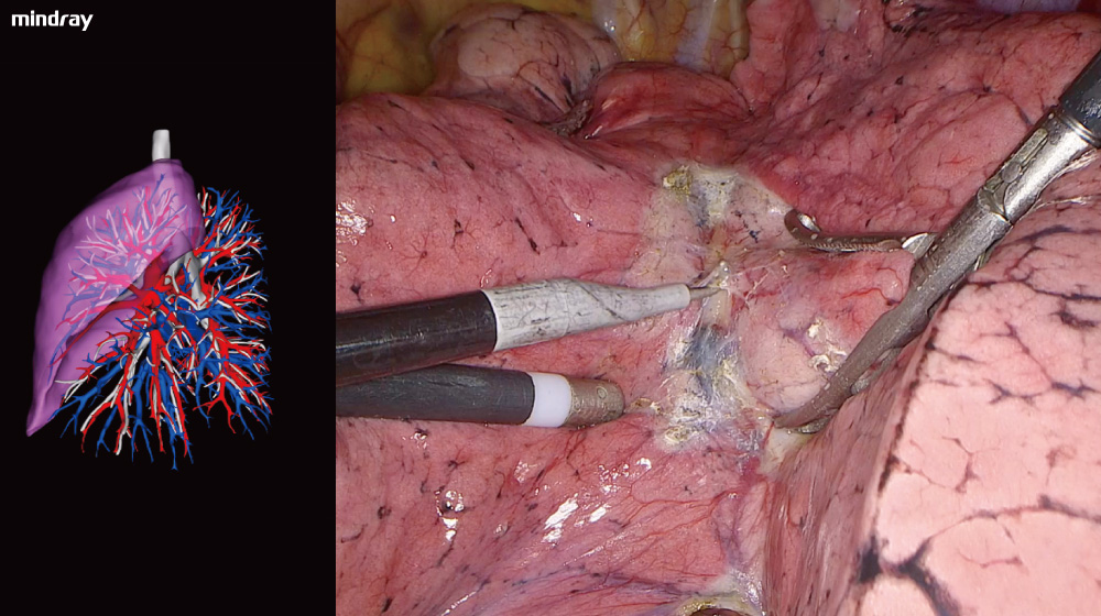
The endoscope can display a 3D reconstruction of the organs' structure, together with the surgical view on the same screen, to assist the surgeon with real-time correction needed during the procedure.
Data Sharing
The patient's vital signs data can be customized and displayed on the endoscope screen, allowing the surgeon to evaluate them timely during surgery. Displaying real-time pressure and flow volume of insufflation enhances surgical safety, efficiency, and education, ultimately contributing to better patient outcomes.


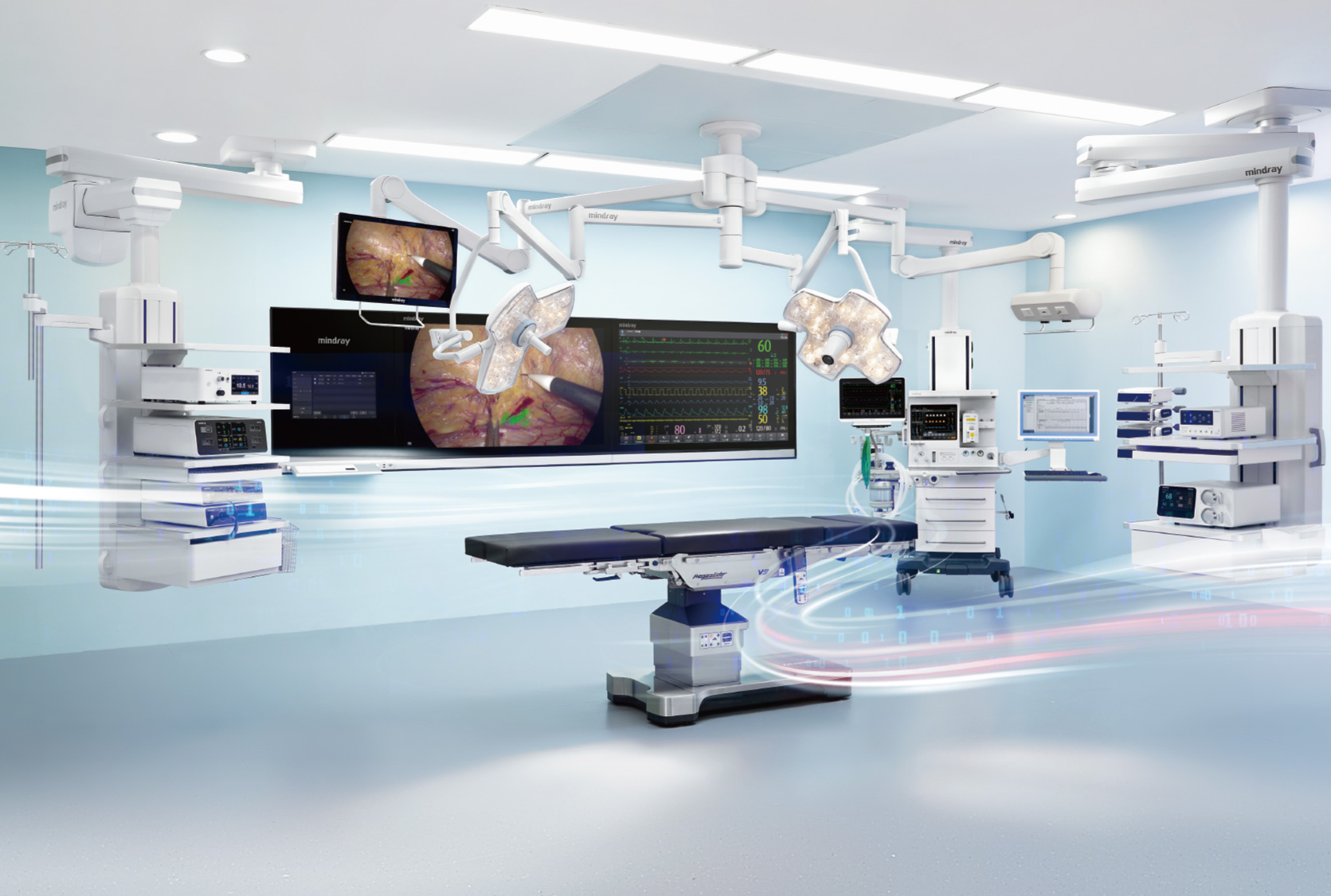
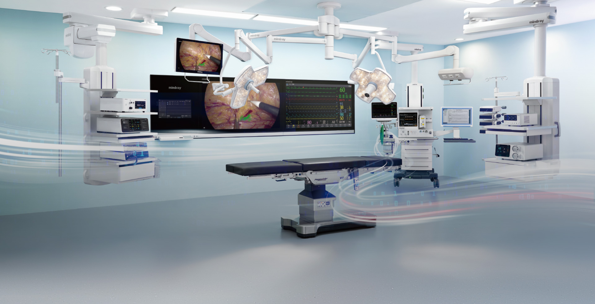
Digital OR, Upgraded Management
The UX7 Endoscope Camera System can be integrated into the Mindray digital operating room for centralized information management, enhancing the overall quality and efficiency of surgical procedures.